

Division of Pathology Ehime Prefectural Central Hospital, Japan
The following detail of movies and images will be published on an American jounal
'Archives of Pathology & Laboratory medicine.'
|
|
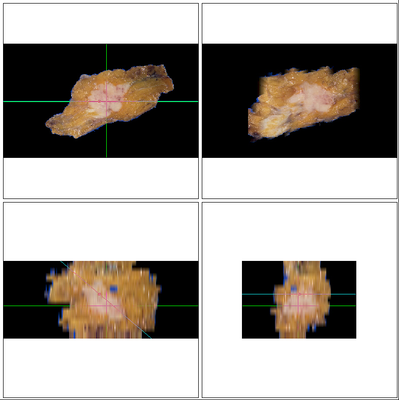 |
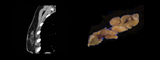
Two-dimensional cut sections of the resected breast specimen : XY(top left), XZ(bottom left), and YX(bottom right) and an arbitrary cut section(top right). | ||
|
|
|
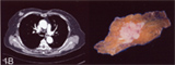
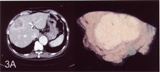
Images from synchronous cut-section movies of enhanced CT image (left) and gross pathologic image (right). | ||
|
|
|
Images from synchronous cut-section movies of enhanced CT image (left) and gross pathologic image (right). | ||
|
|
|
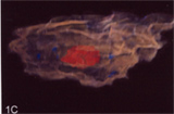
Images from a 3D movie of the resected breast specimen. The red mass is the breast cancer. | ||
|
|
|
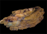
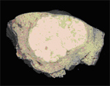
Cut section movie of the gross pathologic images in the arbitrarily cut section plane. In this movie, the cut angle is about 50 degrees. | ||
|
|
|
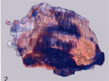
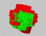
An image from a 3D movie of the resected cardiac myxoma specimen. The left portion was adeherent to the left atrial setum. | ||
|
|
|
The different colors are used to differentiate the myxoma (red) from the non-neoplstic atrial septum (green). | ||
|
|
|
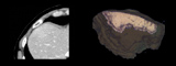
Images from synchronous cut-section movies of enhanced CT image (left) and gross pathologic image (right) of the resected liver specimen. | ||
|
|
|
Images from synchronous cut-section movies of enhanced CT image (left) and gross pathologic image (right) of the resected liver specimen. |
|
|
Cut section movie of the gross pathologic images in the arbitrarily cut section plane. In this movie, the cut angle is about 45 degrees. |